Description
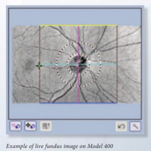
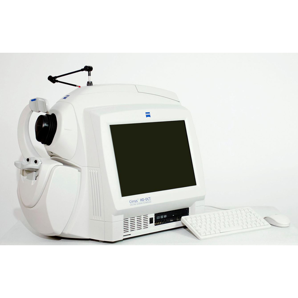
Focused on the essential core OCT functionality, Model 400 is designed with the smaller budget in mind. Live OCT Fundus ™ technology 1 provides the fundus image using the OCT scanner only, rather than an additional line scanning ophthalmoscope (LSO).
Specifications Zeiss Cirrus 400
| Fundus Imaging | |
| Scanning System | Live OCT Fundus Technology |
| Field of View | 36 x 22 degrees |
| OCT Imaging | |
| Methodology | Spectral Domain |
| Scan Speed | 27,000 A-Scans / sec |
| A-scan depth | 2.0 mm (in tissue) |
| Axial resolution | 5 um (in tissue) |
| Transverse resolution | 15 um (in tissue) |
| Computer | |
| Processor | Intel Pentium Core 2 Duo |
| Memory | 2 GB |
| Hard Drive | 750 GB |
| Physical | |
| Size | 26L x 17W x 21H (in) |
| Table Footprint | 39L x 22W (in) |
Specifications Software
- Macular Thickness Analysis
- RNFL Thickness Analysis
- Normative Database – RNFL
- Advanced Visualization and 3D Display
- Anterior Segment Imaging
- 5-line Raster Scan
- Compatible with Cirrus Review Software (license sold separately)
- Connectivity via ZEISS DICOm Gateway
- Optic Nerve Head (ONH) Analysis
- Normative Database – ONH
- Guided Progression Analysis (GPA)
- Macular Change Analysis
- Normative Database – Macula

