You have until Dec 31, 2016 to Write Off the full amount your equipment purchase!
Here is a snapshot of this Tax Code you can take advantage NOW
Here is a snapshot of this Tax Code you can take advantage NOW
Portable slit lamps have come a long way in the last few years. They are no longer manufactured from low-quality plastic and lenses.
Now, you will find a refined, durable, and high-quality lens in its place with options for LED illumination or even full-color screens.
Illumination control and slit width are standard, and they weigh almost nothing. Perfect for applications in remote areas or bed patients and children.
Here we examine three options you should consider for your next hand-held slit lamp. Continue reading 3 Alternatives for Your Next Portable Slit Lamp Purchase
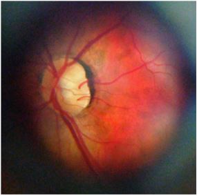
D-EYE, a leading developer of advanced devices for mass health screenings and data analytics is pleased to share the publication of a study comparing the D-EYE Ophthalmoscope with slit lamp technology.
Continue reading D-Eye Ophthalmoscope Scores Well for Glaucoma Screening
“The D-EYE Retinal Imaging System efficiently captures quality photos of the posterior pole in situations where standard fundus photography was impractical” – Doctor Pihlblad
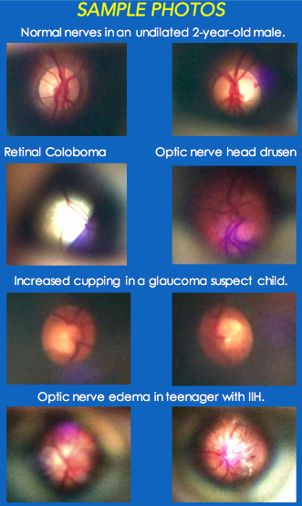
PADOVA, Italy and PASADENA, Calif. – May 10, 2016– Matthew S. Pihlblad, MD and Steven G. Stockslager, MD, physicians from the Ross Eye Institute, Department of Ophthalmology, The State University of New York at Buffalo incorporated the D-EYE Smartphone Retinal Imaging System into a recent study to determine if the system was a beneficial alternative for fundus imaging in the pediatric population. The results of the study were presented to the American Association of Pediatric Ophthalmology (AAPOS) at the organization’s annual conference.
Continue reading D-eye Ophthalmoscope for Pediatric Examination – Case Study

The new D-Eye Ophthalmoscope , designed by Doctor Andrea Russo, allows to perform a screening of the retina through an optical device that attaches magnetically to the back camera of the most popular smartphones.
This allows to get high resolution pictures and video of the retina for further diagnosis. It includes a free App (iphone and android) that will guide the doctor step by step how to take the pictures. The App will store and share the patient’s information. Continue reading D-Eye Ophthalmoscope: Interview with the inventor, Dr. Andrea Russo
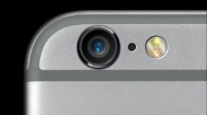
An intelligent way to take excellent slit lamp pictures is with an iPhone slit lamp adapter.
If you are a curious person, then by now, you have been trying to take pictures through your slit lamp with your iPhone, right?
I will detail the advantages of using an iPhone slit lamp adapter as your primary tool for digital photography, the different options you will find in the market, and how to use it to create fabulous pictures.
What did you try to do when you got your first iPhone? For me, it was the camera. The TV ads would not stop promoting the idea that this new iPhone would be the only camera we needed, so I had to test it—and I was not disappointed.
While Apple kept busy launching new versions of the iPhone, 3, 4, 4s, 5, 5s, etc., the camera has always been one of the main focuses of improvement: from the starting 3 megapixels on the first version to today’s 8 megapixels, bigger sensors, improved focus, and faster pictures.
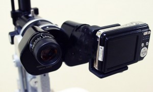
A few years ago
, the optometrist took pictures of the anterior segment using a simple “point and shoot” camera and focused through the slit lamp eyepiece.
Today we have a slit lamp camera adapter that will plug in directly over the eyepiece or replace it. These compact cameras will also allow you to take videos useful for fundus photography.
We might think that due to the growing popularity and quality improvements on smartphone cameras, they are the best option for slit lamp examination. Still, most of the time, it will be faster and more convenient to dedicate a compact camera.
Continue reading Slit Lamp Camera Adapter – A Solid Option for Eye Photography
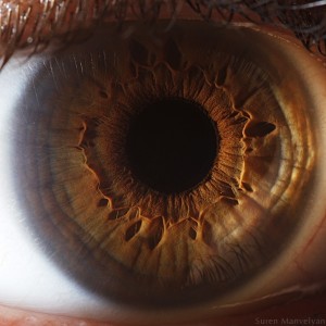
I can still remember the first time I used a slit lamp.
After looking at it and thinking, “What a weird shape for a microscope,” I sat on the stool and looked through the eyepieces into a human eye like I’d never looked before.
My first thought was, “Wow!“. That was it. I would never see the eyes in the same way again. I had discovered the beauty and detail of the fantastic EYE.